
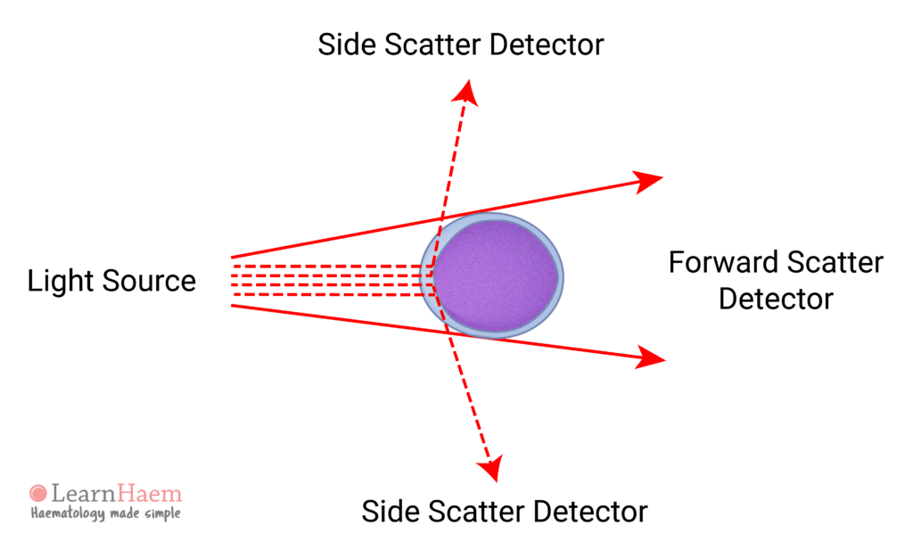
As light enters a cell, the laser beam is scattered in angles and forwards. The amount of light scattered depends on the size of the cell and on the internal complexity (e.g. nuclear complexity, cyotoplasm granularity) of the cell. The light source and the scattered light signals are of the same wavelength, and can hence be determined without specific fluorescence probes.
Forward Scatter
Light defracted at narrow angles is called the forward scatter. The forward scatter is proportional to the size of the cell.
Side Scatter
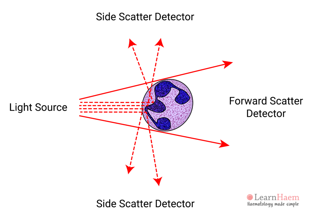
Light can be deflected by obstacles (e.g. cytoplasmic granules or the cell nucleus) within the cells. This scatter is detected by photodiodes positioned at right angles (90°) to the original light source, and is known as side scatter. The side scatter is proportional to the granularity of the cell. In the example below, neutrophils have many cytoplasmic granules. Light is refracted as it hits each granule, causing a high side scatter.
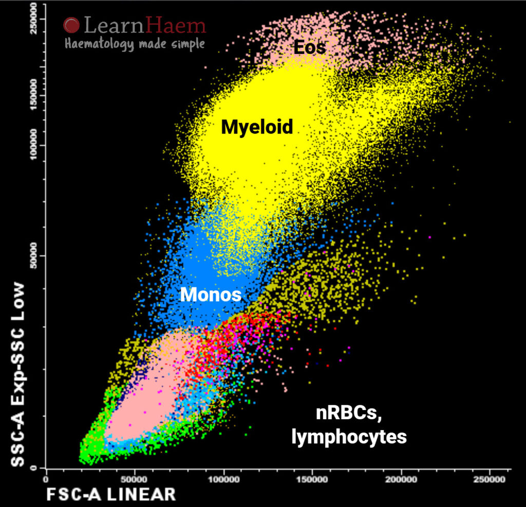
Side Scatter / Forward Scatter Plots
SSc and FSc signals can be interpreted in conjunction to allow cells to be differentiated.
Leave A Comment