
Question 1
A 50-year-old male is referred for anaemia. Hb 11.1 TW 8.9 Plt 264.Toolbar
Adjustments
Brightness
Contrast
Saturation


Report the peripheral blood film. You can adjust the brightness using the settings button in the top right hand corner.
Numerous elliptocytes with some acanthocytes and Howell-Joly bodies. Occasional target cells and spherocytes. Platelet anisocytosis with occasional large platelets seen.
Give three causes for the features seen on the blood film. What is the diagnosis?
Diagnosis: hereditary elliptocytosis post-splenectomy. Other possibilities include: iron deficiency anaemia (less likely as not microcytic or hypochromic), or myelodysplastic syndrome (no dysplasia in the other cell lines).
How would you manage this patient?
Get a family history of hereditary spherocytosis and find out if a splenectomy was done previously. If there is a family history with compatible morphology, no further investigation is required.
Question 2
A 63-year-old male is referred for leukocytosis. Hb 12.1 TW 28 Plt 27.Toolbar
Adjustments
Brightness
Contrast
Saturation
List 5 important features on the blood film.
- Atypical lymphoid cells – mature with no nucleoli, fimbriated cytoplasm
- No monocytopaenia
- Genuine thrombocytopaenia
- No significant red cell fragmentation, mild rouleux
- No large atypical lymphoid cells
Give three possible differential diagnoses, in order of likelihood.
- SMZL
- Hairy cell leukaemia
- Variant hairy cell leukaemia
A bone marrow aspirate and trephine biopsy is ordered. Give three specific investigations which would help confirm your suspected diagnosis.
- Immunophenotyping – look for a non-specific immunophenotype, 5-/11c-/25-/103-
- Annexin A1
- BRAF mutation status
Question 3
A 60-year-old male is incidentally found to have lymphadenopathy on a CT scan done for trauma assessment. Hb 12.1 TW 10 Plt 192.
Toolbar
Adjustments
Brightness
Contrast
Saturation
Report the peripheral blood film and give 3 differentials.
Mild lymphocytosis with mostly small, mature atypical lymphoid cells. Atypical cells have no nucleoli and occasionally have cleft nuclei. There are no large lymphoid cells. No spherocytes or smudge cells. Platelet count is normal.
Differentials: mantle cell lymphoma, chronic lymphocytic leukaemia, marginal zone lymphoma.
The immunophenotype is CD19+/20+/5+/10-/23-/kappa+/smIgM+/22+. What is the most likely diagnosis?
Mantle cell lymphoma
How would you manage this condition?
Evaluate for symptoms which may be an indication for treatment. These include B symptoms, cytopaenias as a result of BM infiltration, massive lymphadenopathy / hepatosplenomegaly causing compression etc. If there are no symptoms, then a watch-and-wait strategy can be employed. If the patient is symptomatic and fit, then cytarabine-based induction followed by consolidative autologous stem cell transplant is the treatment of choice.
Question 4
A 68-year-old female is referred for thrombocytosis. Hb 13 TW 25 Plt 1881.
Toolbar
Adjustments
Brightness
Contrast
Saturation
List 5 important features on the blood film.
Anisopoikilocytosis with majority microcytic and hypochromic RBCs. The RBC count does not appear to be low. Leukocytosis with no myelocytes, metamyelocytes or circulating blasts. Marked thrombocytosis with platelet anisocytosis and occasional hypogranular platelets.
Give three differentials for the PBF and give the likely unifying diagnosis.
Diagnosis: polycythaemia vera with concurrent iron deficiency.
Differentials: myelofibrosis (less likely as no leukoerythroblastosis), CML
List three investigations that you would do to confirm your diagnosis.
- Jak2 V671F
- Erythropoietin
- Ferritin
How would you manage this patient?
The patient is high risk based on age. She requires aspirin and cytoreduction with hydroxyurea.
Question 5
A 30-year-old female attends her regular Haematology follow-up for review. Hb 9.8 TW 8.4 Plt 203.
Toolbar
Adjustments
Brightness
Contrast
Saturation
List 5 important features on the blood film.
Dimorphic picture. One population is normocytic, normochromic, while the other is microcytic and hypochromic. The microcytic, hypochromic population has numerous target cells, circulating nRBCs, acanthocytes and Howell-Joly bodies. Mild platelet anisocytosis.
The patient’s diagnostic HPLC is shown below. Interpret the HPLC. What is the diagnosis?
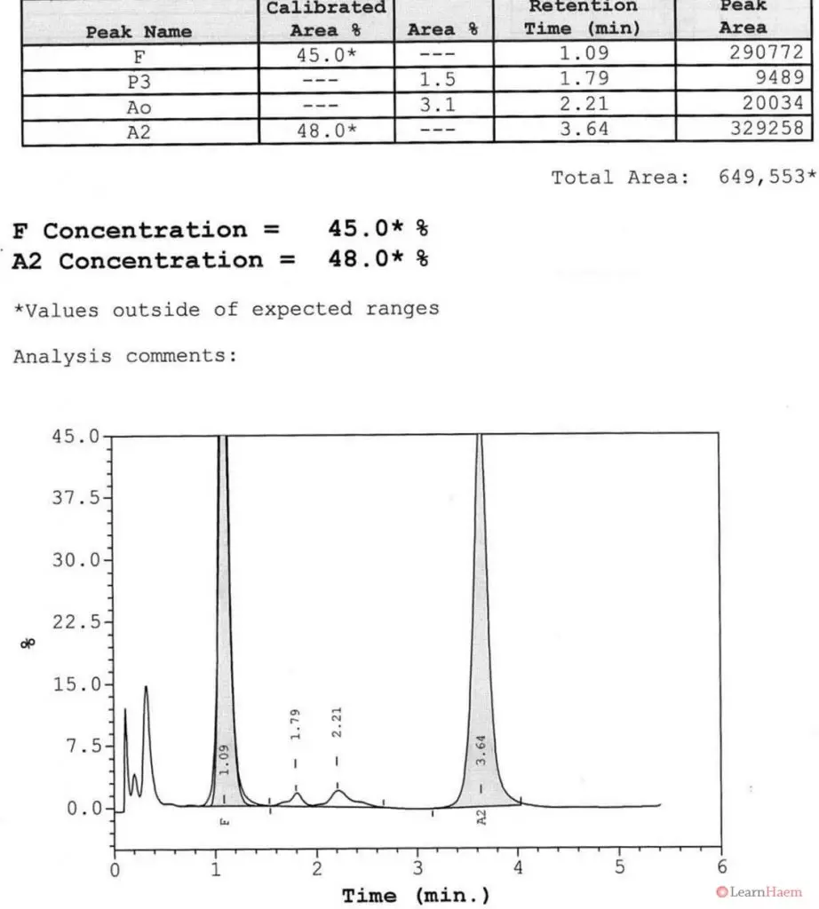
2 major variant haemoglobin with elution times corresponding to HbF and HbA2. The variant haemoglobin in the A2 window is likely to be HbE. Given that there is no HbA, this suggests that the diagnosis is a compound heterozygote for HbE/β0 thalassaemia.
Give three features on the PBF which are unexpected given the diagnosis suggested by the HPLC. What possible reasons are there to explain these features?
The dimorphic picture, presence of acathocytes and Howell-Joly bodies. Possible explanations include recent transfusion and post-splenectomy.
What an incredible tool! I”m amazed by its complexity and efficiency! Thank you guys for this,
NICE…GREAT EFFORTS .