
Indications for splenectomy
- ITP
- AIHA
- Thalassaemia intermedia (old indication)
- Congenital RBC membrane defects
- Hereditary spherocytosis
- Hereditary elliptocytosis
- Lymphoproliferative disorders
- Splenic marginal zone lymphoma
- Hairy cell leukaemia (old indication)
- Myelofibrosis (old indication)
- Trauma

Blood film features:
- Howell-Jolly bodies (nuclear remnants which are usually removed by the spleen)
- Pappenheimer bodies (removed from reticulocytes in spleen)
- Can also be seen in iron-loading anaemias
- Acanthocytes
- Target cells
- Spherocytes
- Stomatocytes
- Thrombocytosis
- Platelet anisocytosis
- Lymphocytosis


The two blood films above are from a patient who had a splenectomy, showing target cells, acanthocytes, Howell-Jolly bodies, basophilic stippling and Pappenheimer bodies.
This is the blood film of a patient who had a splenectomy for trauma, then subsequently developed cancer which metastasized to the bone. The latter explains the numerous nRBCs circulating.
This blood film is from a transfusion-dependent patient who is a compound heterozygote for HbE/beta-thalassaemia. There is a dimorphic picture in the background, with a population of normochromic, normocytic transfused cells, as well as the abnormal native microcytic, hypochromic cells with anisopoikilocytosis and prominent target cells. The splenectomy features can be seen in these cells.
The two slides above are from a patient who had a splenectomy for hereditary spherocytosis. Numerous spherocytes can be seen in the background. There are a number of acanthocytes with prominent Howell-Jolly bodies.
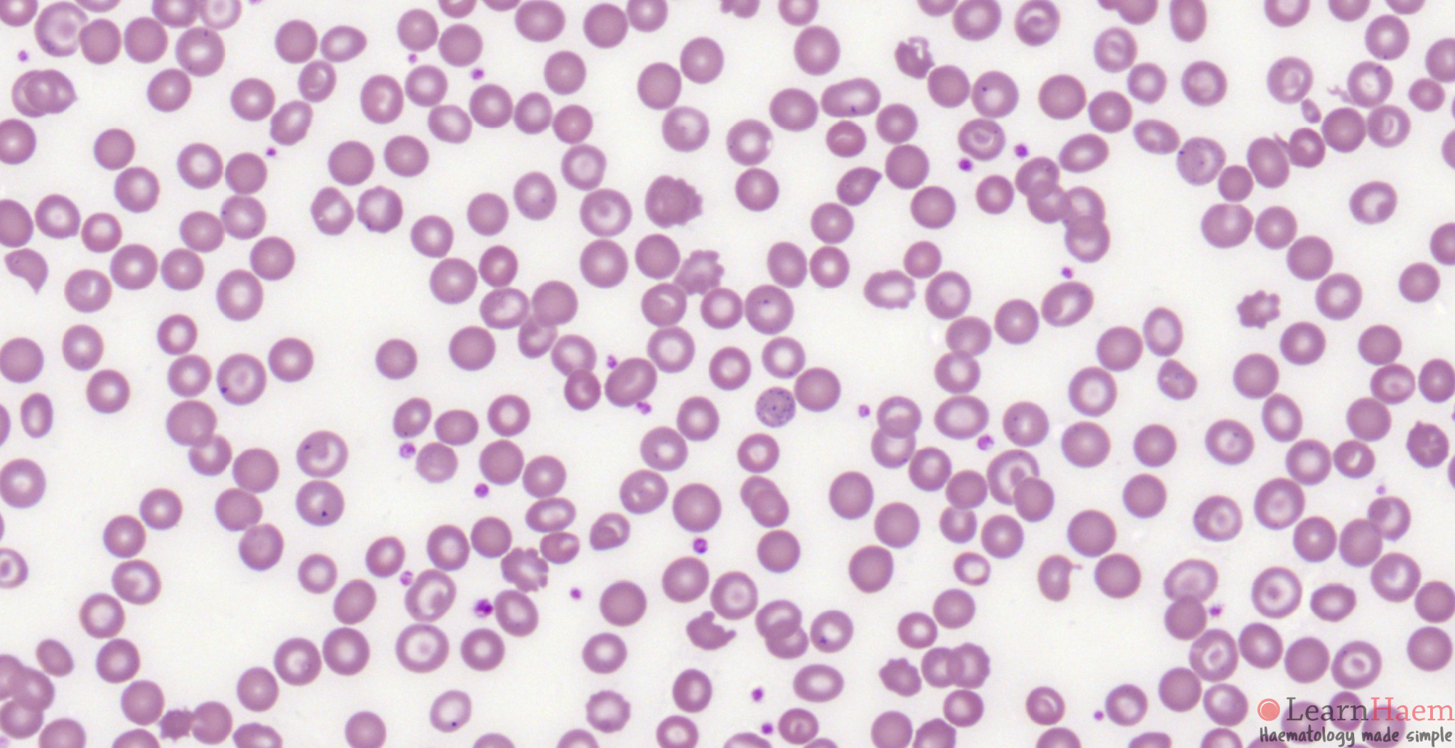
This patient had a splenectomy for hereditary spherocytosis. In contrast to the patient above, there are less acanthocytes. However, Howell-Jolly bodies and Pappenheimer bodies are readily seen. In addition, there is a mild thrombocytosis. The majority of RBCs are spherocytes.
This patient has hereditary elliptocytosis and subsequently underwent a splenectomy. Note the prominent acanthocytes, elliptocytes and contracted elliptocytes. Adjust the brightness using the toolbar if the image appears too dark.
This patient underwent a splenectomy for splenic marginal zone lymphoma. The blood film was made >20 years after the splenectomy, when the patient was in remission.
Other features to look for:
- Features of the underlying disease
- Spherocytes – hereditary spherocytosis
- Target cells – thalassaemia
- Atypical lymphoid cells – lymphoproliferative disorders
- Normal blood film – consider splenectomy post-trauma or for ITP
Thank you so much. I’m in California (Bay Area) preparing for the ASCP (MLT) exam !!!!